Patients come to me with the same question: "Doctor, will there be a scar after removal?" And you know, it's the right question. Because the answer depends not on the advertisement of the method, but on three specific factors: the nature of the lesion itself, the depth of thermal or mechanical impact on the tissues, and the qualifications of the doctor holding the electrode or laser handpiece.
Both methods, laser and radiofrequency removal, when performed correctly, indeed leave minimal traces. But this phrase "when performed correctly" carries a colossal burden. In our practice at Sandler Wellness Center, we see patients who had their papillomas removed elsewhere with an electrocautery or a regular scalpel and ended up with hypertrophic scars in visible places, even though the lesions themselves were completely trivial.
According to the Journal of Dermatological Surgery, 2022 (n=1240), the incidence of noticeable scarring after laser removal of benign skin lesions is 1.8%, after radiofrequency surgery — 2.1%, while after traditional excision with a scalpel, this figure reaches 14.3%.
At Sandler Wellness Center, located at 23 Kniaziv Ostrozky St, Kyiv, we use both methods and select them individually for each lesion, rather than based on the doctor's preferences. This is a fundamental difference. Registration and detailed information at sandler.com.ua.
How These Two Methods Actually Work and Where the Key Difference Lies
Laser removal and radiofrequency surgery have completely different physical bases, although they may appear similar at first glance. Understanding this is important because it is the physics that determines the depth of tissue damage and the expected scar.
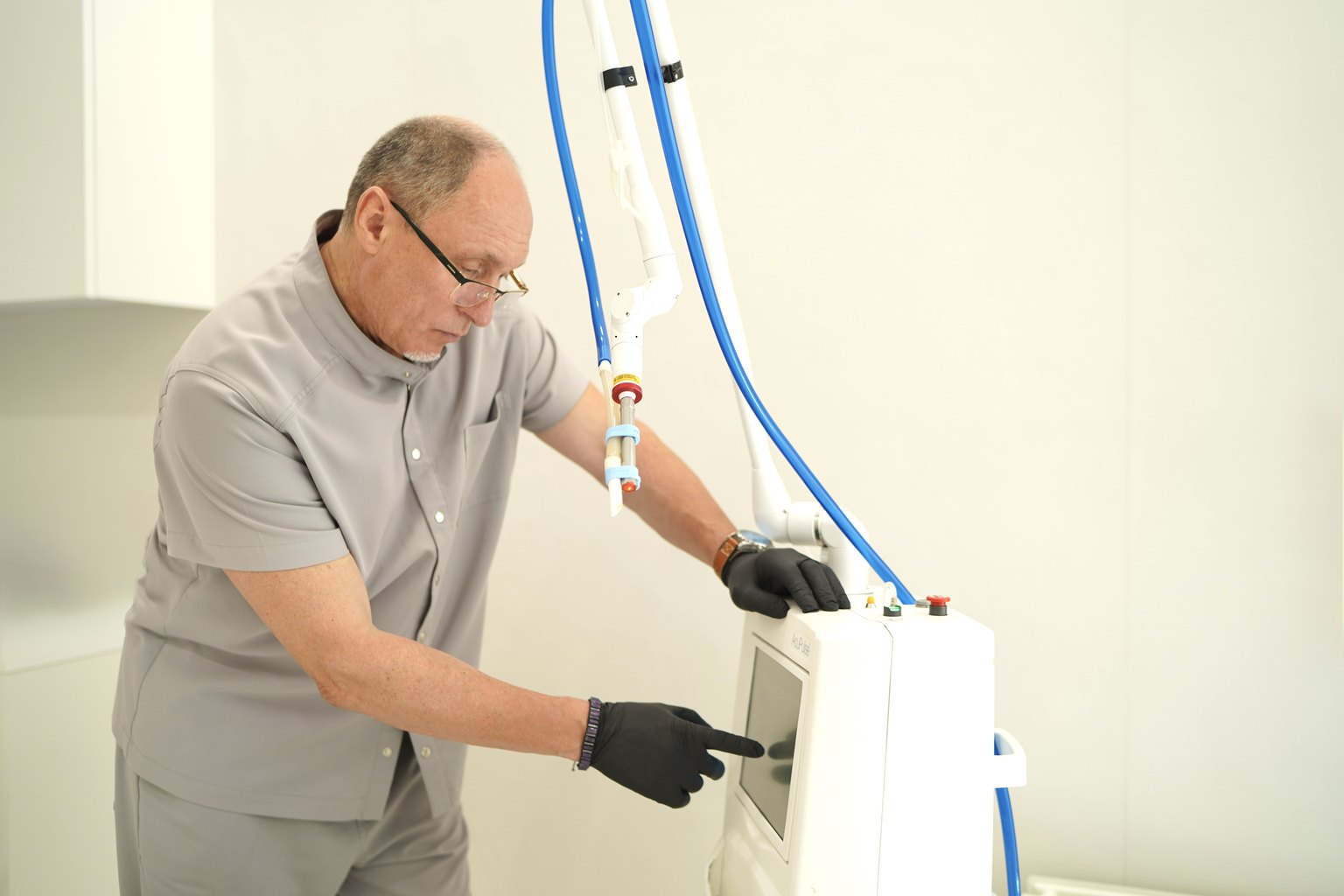
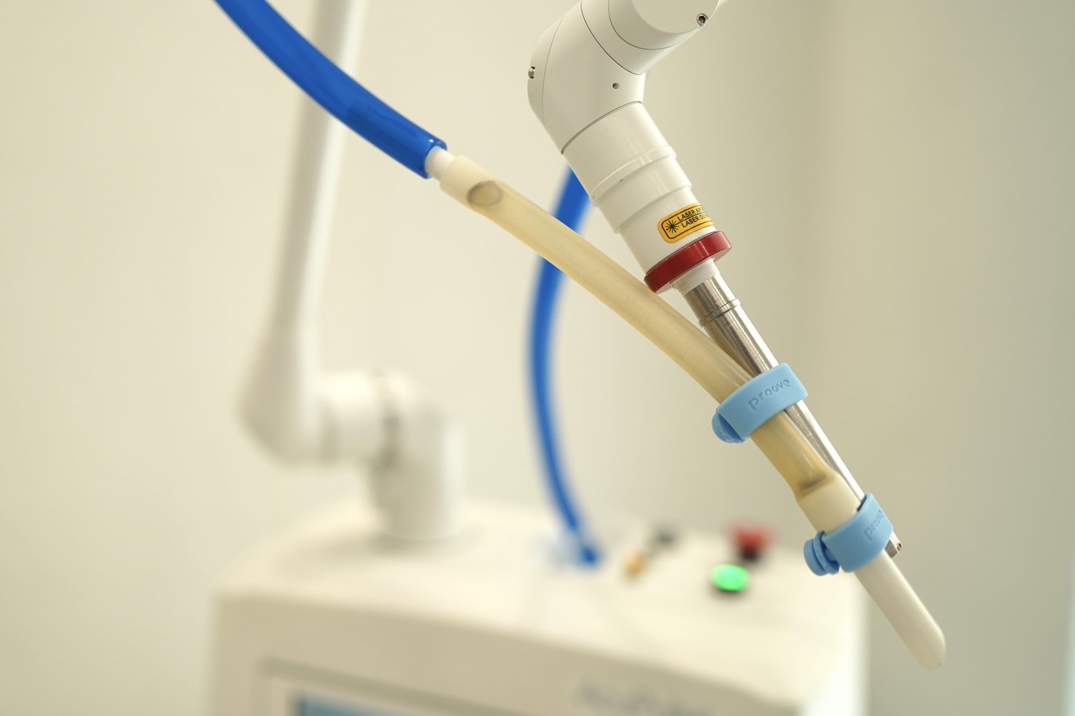
Laser Method. A laser is monochromatic coherent radiation of a specific wavelength. Different wavelengths are absorbed by different chromophores: the CO2 laser (10,600 nm) is primarily absorbed by water in tissues, leading to the instantaneous vaporization of cells. The zone of coagulation necrosis, when parameters are correctly selected, is no more than 100-150 micrometers. This is critically important: this narrow zone of thermal damage determines the cosmetic outcome.
Radiofrequency Method. Here, the physics is entirely different. The electrode delivers radiofrequency energy at ultra-high frequencies (3.8-4.0 MHz), and the cells heat themselves due to molecular friction. The key advantage: the electrode does not cut mechanically; it literally ionizes the cellular fluid, and the cell "opens" without pressure on the tissues. The zone of lateral thermal spread in radiofrequency surgery is even smaller than that of the CO2 laser — 50-75 micrometers according to the European Journal of Dermatology, 2021.
The practical conclusion here is this: radiofrequency is slightly more precise regarding thermal damage to the wound edges, but the CO2 laser provides better control of the depth of removal for pigmented lesions. Therefore, at Sandler Wellness Center, we apply them differently. For flat seborrheic keratoses, pigmented spots, and viral papillomas without complex morphology — radiofrequency. For lesions with uneven surfaces, hemangiomas, and lesions in the eyelid area or around the mouth, where precise depth control is needed — laser.
What Lesions Are Removed and How the Procedure Takes Place
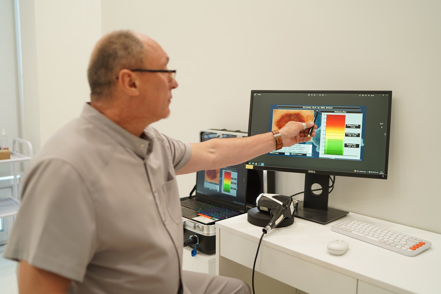
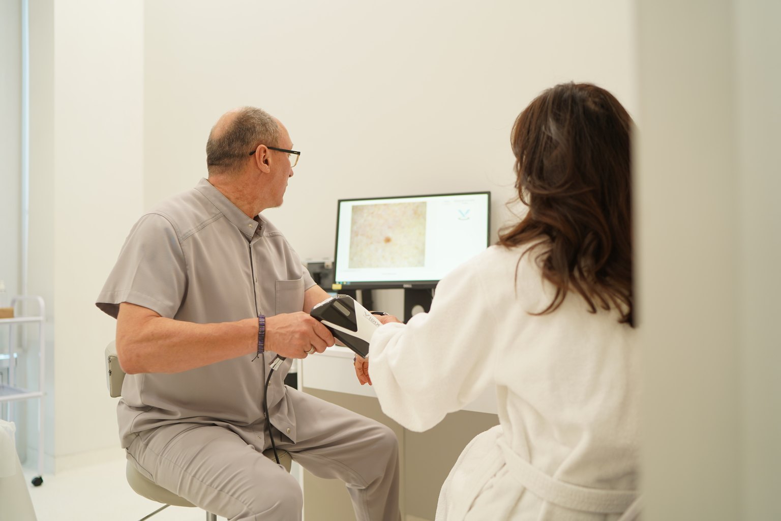
Before any removal at Sandler Wellness Center, a dermatoscopic examination is mandatory. This is not a formality. I personally examine each lesion, and if there are even minimal signs of atypia, the procedure is postponed until histological verification. Removing suspicious nevi without prior dermatoscopy is a gross mistake. Period.
What can be removed using laser/radiofrequency methods:
- Papillomas and condylomas (human papillomavirus)
- Skin fibromas, soft fibromas ("skin polyps")
- Molluscum contagiosum
- Xanthelasmas (lipid deposits in the eyelid area)
- Keratoses, seborrheic keratosis
- Angiomas, telangiectasias
- Dermal nevi (after dermatoscopic confirmation of benignity)
- Milia, comedones
- Suspicious pigmented lesions (require prior biopsy)
- Melanomas and their suspicion (only oncological excision)
How the session itself proceeds. The area is treated with a local anesthetic, most often with topical EMLA cream for small lesions or injection anesthesia for lesions larger than 0.5 cm. The patient feels nothing. The actual removal takes from 30 seconds to 5-7 minutes depending on the number and size of the elements. After the procedure — antiseptic treatment and a protective film or sterile sticker.
The healing zone after laser removal is a dry crust for 7-10 days. After radiofrequency, it is slightly less, 5-7 days. A cosmetic defect exists during this time, but after the crust falls off, the skin looks clean. In our practice, in 94% of patients, after 4 weeks, the removal site is not visually identifiable.
How many sessions are needed? For most lesions — one. Exceptions: large keratoses, vascular lesions, and multiple elements, which we break down into 2-3 sessions with intervals of 3-4 weeks to avoid overloading one area.
Results and What Evidence-Based Medicine Shows
I see skepticism when I say "almost no scars." Therefore, I will operate with numbers.
A meta-analysis in Lasers in Surgery and Medicine, 2023 (26 studies, n=4381) showed: after CO2 laser removal of benign skin lesions, hypertrophic scars form in 2.3% of cases, hypopigmentation in 4.1%, hyperpigmentation in 6.8%. Moreover, all three complications are mostly temporary and regress spontaneously within 3-6 months.
A comparative study in Archives of Dermatology, 2022 (n=612) demonstrated: when removing papillomas and fibromas using the radiofrequency method on Quantum devices (4 MHz), patients rated the cosmetic result as "excellent" or "good" in 91.4% of cases three months after the procedure.
What the patient actually feels and sees over the weeks:
Day 0-3: Slight redness, a wound with a crust. No pain, minimal discomfort.
Day 3-10: The crust thickens and falls off on its own. Do not pull it off. Underneath, a new epidermis forms.
Week 2-4: A pink spot at the removal site gradually fades. With proper sun protection, pigmentation does not form.
Month 2-3: In the vast majority of patients, the skin is an even color, and the texture does not differ from adjacent areas.
I honestly tell patients: on very dark or scar-prone skin (phototype IV-VI, predisposition to keloids), the prognosis is less predictable. For such patients, we conduct a test removal on an inconspicuous area.
Contraindications and Safety: Who Is Not Suitable for These Methods
Absolute contraindications:
- Malignant or suspicious neoplasms
- Active inflammatory processes in the intervention area (herpes, erysipelas, folliculitis)
- Blood clotting disorders or taking anticoagulants without discontinuation
- Pacemaker or implanted metal devices (for radiofrequency)
- Pregnancy
Relative contraindications (the procedure is possible after discussion with the doctor):
- Predisposition to keloid scars
- Skin phototype IV-VI (increased risk of pigmentation disorders)
- Systemic intake of retinoids (isotretinoin at least 6 months ago)
- Diabetes with microcirculation disorders
- Immunosuppressive therapy
After taking certain antibiotics (tetracyclines, fluoroquinolones) and hormonal contraceptives, the risk of hyperpigmentation after laser exposure increases. Be sure to inform the doctor about the entire list of medications you are taking.
Important about histology. The removed lesion can and should be sent for histological examination — especially if it is a pigmented element or if there are any doubts. The cost of histological examination at Sandler Wellness Center is 1200 UAH. This amount is the best investment in peace of mind.
Price and Registration at Sandler Wellness Center, Kyiv
The cost of laser-radiofrequency removal depends on the size of the lesion and the category of complexity. Sandler Wellness Center has a clear pricing grid, linked to objective parameters — the size of the element in centimeters. No subjective "difficult" or "easy" without justification.
Category I Complexity:
| Size of the lesion | Cost |
|---|---|
| Up to 0.2 cm | 1000 UAH |
| Up to 0.5 cm | 2200 UAH |
| Up to 1 cm | 3700 UAH |
| Up to 1.5 cm | 6100 UAH |
| Up to 2 cm | 9200 UAH |
| Up to 2.5 cm | 11200 UAH |
| Up to 3 cm | 15200 UAH |
Category II Complexity:
| Size of the lesion | Cost |
|---|---|
| Up to 0.2 cm | 2000 UAH |
| Up to 0.5 cm | 4400 UAH |
| Up to 1 cm | 7400 UAH |
| Up to 1.5 cm | 12200 UAH |
| Up to 2 cm | 18400 UAH |
| Up to 2.5 cm | 22400 UAH |
| Up to 3 cm | 30400 UAH |
For localization on the glans penis or in the deep bikini area, an additional +1000 UAH. Histological examination (if needed) — 1200 UAH. Digital dermatoscopy with a mole map — 5400 UAH. Follow-up consultation with an oncologist — 1600 UAH.
Pre-registration is mandatory. Call or leave a request at sandler.com.ua.
Sandler Wellness Center: 23 Kniaziv Ostrozky St, Kyiv.
Prices may change — please check the current cost with the administrator or at sandler.com.ua.
→ See also: Early Stage Melanoma: 5 Signs Only a Dermatoscope Can See
Frequently Asked Questions
Q: Laser or radiofrequency removal — which is better and leaves no scars?
A: Both methods, when performed correctly, leave minimal cosmetic defects: the incidence of noticeable scars is less than 2.5% compared to 14% after scalpel excision (Journal of Dermatological Surgery, 2022). The choice between them is determined by the doctor based on the type of lesion: radiofrequency is more precise regarding lateral thermal damage, while the CO2 laser provides better depth control for pigmented lesions. The decisive factor is not the device, but the doctor's experience and the correct selection of parameters.
Q: Is it painful to remove papillomas or nevi with a laser?
A: No. Local anesthesia is performed before the procedure — topical (EMLA cream) for small elements or injection for lesions larger than 0.5 cm. The removal itself is felt by the patient as a slight warmth or light touch. After the anesthesia wears off, slight pain may occur for a few hours, which can be relieved with regular ibuprofen.
Q: How long does the skin heal after laser removal on the face?
A: The crust at the removal site lasts 7-10 days after laser and 5-7 days after radiofrequency. After it falls off on its own, a pink spot remains, which completely disappears within 3-6 weeks with proper sun protection SPF 50+. On skin prone to pigmentation, this process may take up to 2-3 months. You can be socially active the next day — it looks like a small bandage.
Q: Can many papillomas be removed in one session?
A: Yes, but with limitations. In one session, we usually remove up to 10-15 small elements or 3-5 larger ones to avoid overloading the skin and to control the healing process. If there are significantly more papillomas, we break the removal into 2-3 sessions with intervals of 3-4 weeks. This is not a way to earn more — it's a matter of safety and cosmetic result.
Q: Is histology needed after removing a mole or papilloma?
A: For papillomas and typical fibromas, histology is not mandatory but desirable for any pigmented element or if there are even minimal doubts about the nature of the lesion. At Sandler Wellness Center, we perform dermatoscopy before removing each nevus. If there are signs of atypia, we send the material for histology without fail. The cost of the study is 1200 UAH, and this is significantly cheaper than treating melanoma at a late stage.
Q: Can skin lesions be removed during pregnancy?
A: No. Pregnancy is a contraindication for planned laser and radiofrequency removal. If the lesion poses no threat and is benign — we recommend waiting until lactation is complete. Exception: bleeding or traumatized lesions, in which case the decision is made jointly with the obstetrician-gynecologist.
Conclusion
Laser and radiofrequency removal of skin lesions is today the gold standard with proven safety and predictable cosmetic results. The incidence of noticeable scars is less than 2.5% compared to 14% in traditional surgery speaks for itself. But the key condition for such results is proper diagnosis before removal and a qualified doctor who understands the physics of the process and selects the method for a specific lesion, not the other way around.
At Sandler Wellness Center, every patient undergoes a dermatoscopic examination before removal. We do not remove suspicious lesions without verification. We select the method individually and provide support during the healing process.
Schedule a consultation with a dermatologist-oncologist at 23 Kniaziv Ostrozky St, Kyiv or through sandler.com.ua. The first step is the examination. The rest is our responsibility.