Every year in Ukraine, over 3,500 new cases of melanoma are registered. Most of these are at a stage when the tumor has already penetrated deeper layers of the skin. And not because people ignore the symptoms. But because the early signs of melanoma are simply not visible to the naked eye.
I am Dmytro Cheshensko, a dermatologist-oncologist and candidate of medical sciences. I see this every week: a person comes in with a "suspicious mole" that they have considered ordinary for months. To the eye — nothing. The dermatoscope shows structures that definitely require removal and histology.
Dermatoscopy increases the accuracy of melanoma diagnosis from 65% (clinical examination) to 90–95% (according to the British Journal of Dermatology, meta-analysis 2022, n=22,000 patients). This is not a marketing figure. This is the difference between "we will monitor" and "we remove it now and save a life."
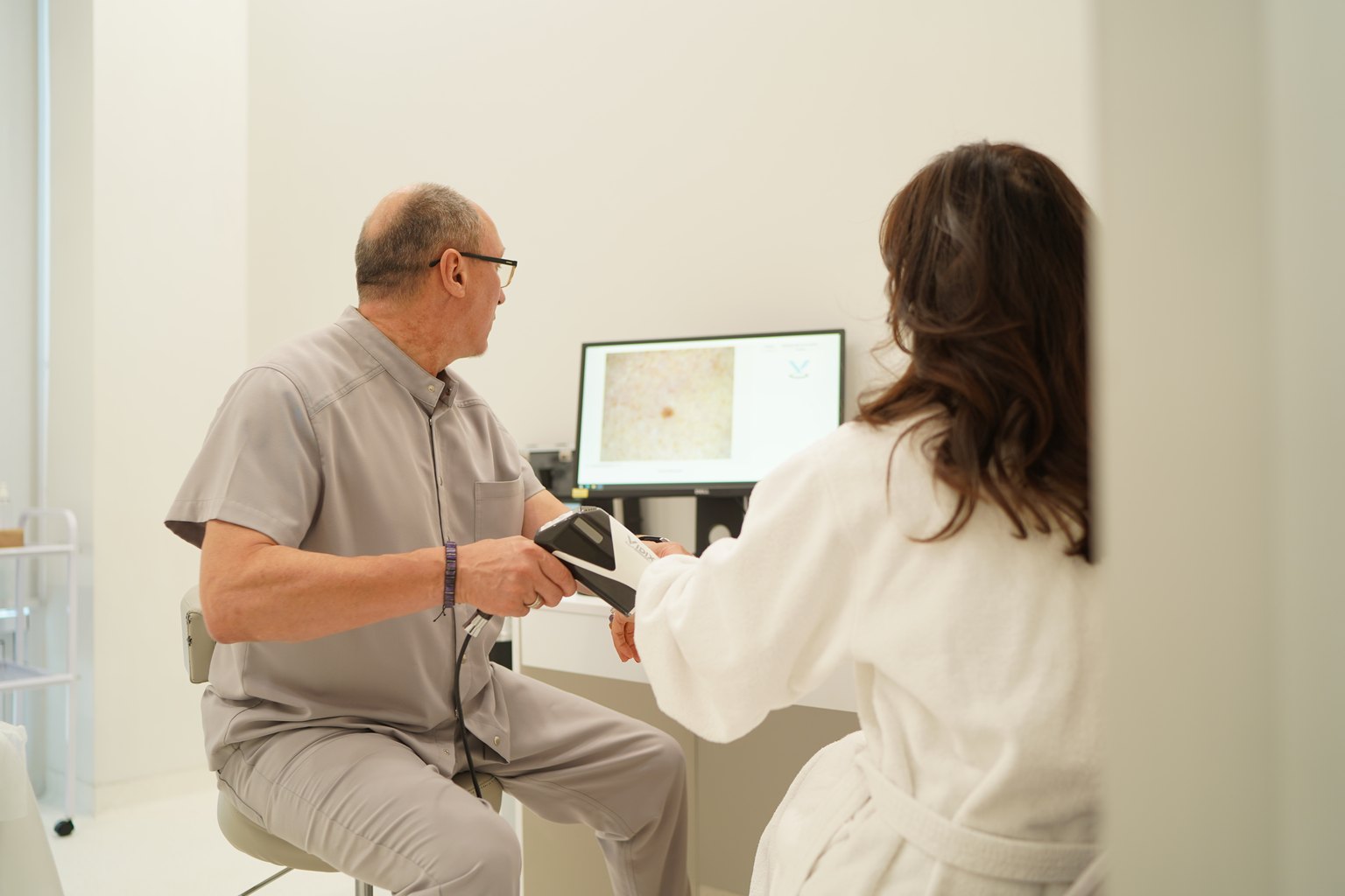
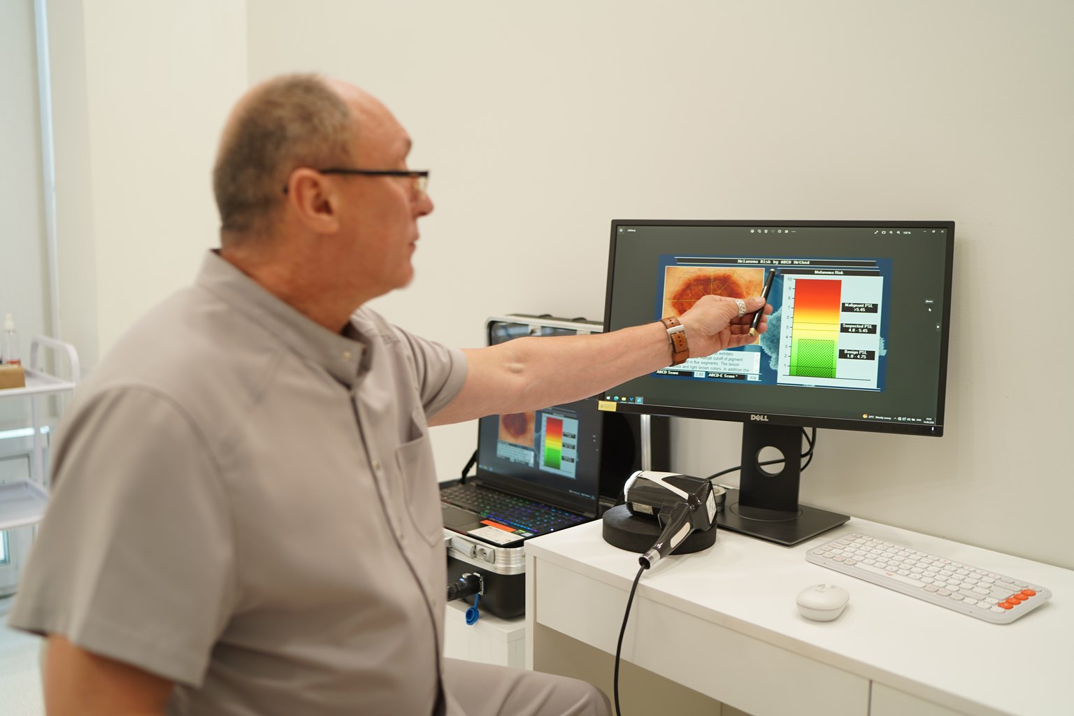
At Sandler Wellness Center located at 23 Kniaziv Ostrozky St, Kyiv, a consultation with an oncologist-dermatologist includes dermatoscopy with the VIDIX device — digital dermatoscopy with image archiving and the ability for dynamic monitoring. If you have even one mole that has changed, this article is for you.
What is Dermatoscopy and Why It Sees What You Don’t
A dermatoscope is a dermatological microscope with polarized or immersion lighting that allows visualization of the structures of the epidermis and the upper layer of the dermis to a depth of up to 0.2 mm. During a regular examination, you see the surface. The dermatoscope shows the architecture.
The human eye distinguishes color and shape. The dermatoscope detects vascular patterns, pigment networks, regressive structures, and atypical globules. This is a different level of information. The analogy is crude but accurate: comparing a clinical examination and dermatoscopy is like comparing a regular photo to an MRI.
What is important to understand about melanoma: it develops horizontally before it starts to grow vertically. The stage of horizontal growth can last from 1 to 10 years. It is during this time that the dermatoscope can detect pathological changes that are not yet on the surface.
The 5-year survival rate for stage I melanoma is 98%. For stage IV, it is 23%. (National Cancer Institute SEER Database, 2023). In simpler terms: melanoma detected in time is a curable tumor. Missed — it is not.
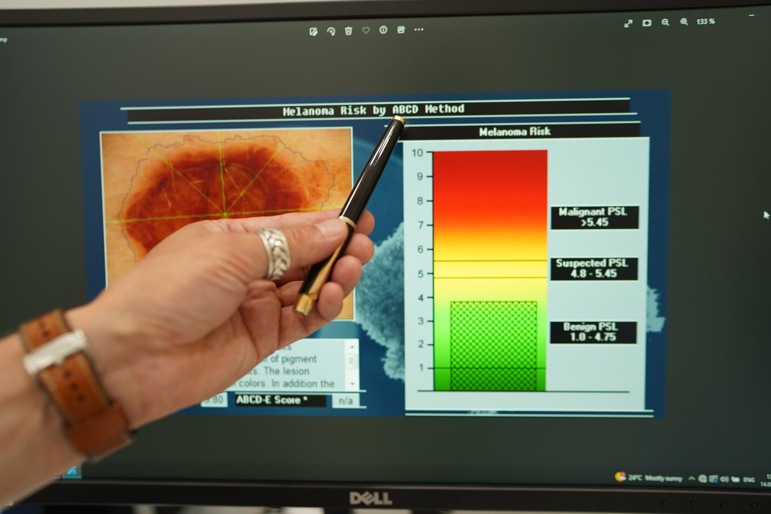
What is the difference between a regular examination and digital dermatoscopy with VIDIX? The VIDIX device used at Sandler Wellness Center stores images of each formation linked to a body map. During a follow-up visit, we compare pixels — literally. Any dynamics in size or structure become visible, even if they are not noticeable to the doctor's eye.
5 Dermatoscopic Signs of Early Melanoma That Only the Device Can See
Here are the specifics. Not general words about "color change" — but what exactly a dermatologist-oncologist looks for in the device's eyepiece.
1. Atypical Pigment Network
A normal mole has a uniform pigment network with similar cells. In melanoma, the network becomes uneven: the thickness of the lines varies, the cells are of different sizes, and the network breaks unevenly at the edges. This is the first and most common sign. According to the Journal of the American Academy of Dermatology (2023, n=3,200), an atypical network is present in 71% of melanomas at the in situ stage.
2. Regression Structures
Whitish-gray areas without a clear structure inside the pigmented formation. These are areas where tumor cells are already destroying normal tissue, and the immune system is trying to fight them. They appear as "scar-like" white areas or a gray-blue veil. Patients sometimes notice that "the mole has lightened a bit" — and consider it a good sign. In fact, it is the opposite.
3. Atypical Vascular Patterns
New vessels that the tumor builds for nourishment (angiogenesis) have a characteristic appearance: twisted, irregular, with dot-like or needle-like loops. In early melanoma, they appear even before the tumor becomes noticeably raised.
4. Blue-White Veil
A diffuse gray-blue zone over areas with dense tumor cells. This is an optical phenomenon: pigment in deeper layers of the dermis combined with hyperkeratosis gives a characteristic coloration. A classic sign of vertical growth — when it is just beginning.
5. Peripheral Pseudopods and Radial Streaming
Finger-like or ray-like extensions of pigment at the edges of the formation. This is the tumor "spreading" across the basal layer of the epidermis. Visible only under the dermatoscope. To the eye, it seems that the edges are just a bit uneven.
Who Needs Dermatoscopy: Indications and Examination Protocol
The short answer: everyone. The long answer — there are high-risk groups where examination is mandatory at least once a year.
Risk Factor Checklist:
- More than 50 pigmented formations on the body
- History of dysplastic nevi (atypical moles)
- First-degree relatives who have had melanoma
- Sunburns in childhood or adolescence (especially blistering)
- Regular visits to tanning beds (any — risk increases by 59%, according to IARC, 2023)
- Fair skin, red or light blonde hair (phototype I–II)
- Work outdoors (farmers, builders, athletes)
- Immunosuppression (post-transplant, during treatment)
- A mole that has changed in size, shape, or color in the last 6 months — this is already a reason to come in today
How does a consultation at Sandler Wellness Center proceed? The appointment takes 30–40 minutes. First, we gather the history: family history, past burns, any changes. Then we examine the entire skin surface — not just the formations that concern you. Melanoma can occur anywhere, including the soles, scalp, and nail beds. Each suspicious formation is photographed with the VIDIX device and described in the protocol.
How many sessions are needed? The initial consultation is one visit. If there are many formations or suspicious findings, a follow-up dermatoscopy is scheduled in 3 months. If suspicion is found — a referral for excision and histology is provided on the same day.
What Research Shows: Real Data on the Effectiveness of Early Diagnosis
Melanoma detected at the in situ or IA stage is treated with surgical removal. Without chemotherapy. Without immunotherapy. Without radiation. Just surgery and monitoring.
Data from the Surveillance, Epidemiology, and End Results Program (SEER, National Cancer Institute, 2023) based on a sample of 124,000 patients: the 5-year survival rate at stage 0 (in situ) is 99.9%, stage IA — 97.6%, stage IB — 92.7%. At stage IIIC, it drops to 40–45%.
A study by the University of Queensland (2022, n=1,847) showed that the implementation of systematic dermatoscopic screening reduced the proportion of melanomas detected at stages III–IV by 34% over 5 years. Not "helped." Reduced by a third.
In our practice, most melanomas we detect are stage IA or in situ. People who have never undergone dermatoscopy before sometimes come in with more advanced processes. The difference is obvious.
What to realistically expect from the examination: if the formation is benign — you receive reassurance and a protocol with photos for comparison at the next appointment. If suspicious — a clear recommendation for removal with specific timelines. No "let's monitor for a year without documentation."
Contraindications and Limitations of Dermatoscopy
Honestly: dermatoscopy has no medical contraindications as a method. It is a non-invasive, painless procedure with no impact on tissues.
However, there are important limitations to be aware of.
What Dermatoscopy Cannot Do:
Even the best dermatoscope and the most experienced doctor cannot replace histology. Dermatoscopy is a diagnosis of probability. The final diagnosis of melanoma or its absence is made only by a pathomorphologist after examining the removed tissue.
There are also situational limitations. Formations under the nail or on mucous membranes may be technically difficult to access with a standard dermatoscope. Nodular melanomas — the most aggressive subtype — sometimes grow so quickly that dermatoscopic criteria lag behind the clinical picture. Therefore, we always evaluate the clinical picture in parallel.
Who does not need dermatoscopy? Theoretically — no one. Practically: if you have no pigmented formations, very dark skin (phototype VI) without risk factors and no symptoms, an examination is still useful but less of a priority. Although melanoma can occur on dark skin — more often on acral areas (palms, soles, nails).
→ See also: Laser and Radiofrequency Removal of Skin Lesions: What Truly Leaves No Scars
Price and Appointment at Sandler Wellness Center, Kyiv
A consultation with an oncologist-dermatologist with VIDIX dermatoscopy at Sandler Wellness Center costs 3,500 UAH. This amount includes: history taking, examination of the entire skin surface, digital dermatoscopy of suspicious formations with photographic documentation, examination protocol, recommendations.
A follow-up consultation with control dermatoscopy costs 1,600 UAH.
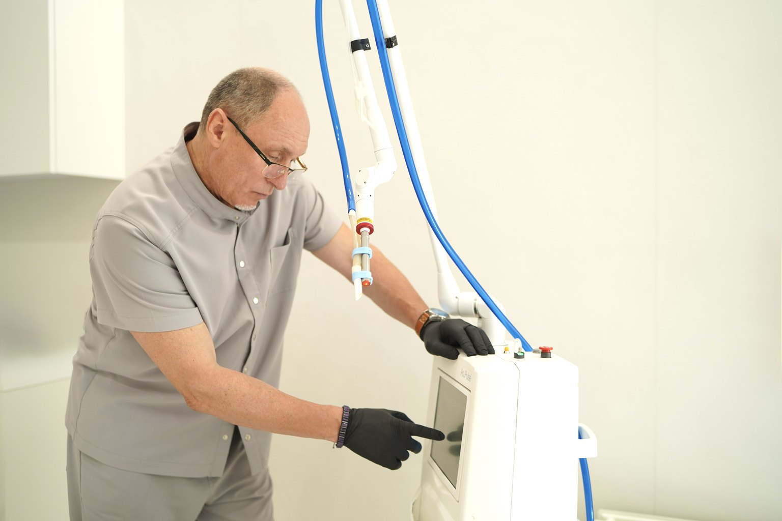
If removal of a formation is needed based on the examination results — laser-radio wave removal of skin formations costs from 1,000 to 30,400 UAH depending on size, quantity, and location. The exact cost is calculated after the examination when we know what and where needs to be removed.
Histological examination of the removed material costs 1,200 UAH.
You can make an appointment through the website sandler.com.ua or by phone. Sandler Wellness Center is located at 23 Kniaziv Ostrozky St, Kyiv. Usually, the first available appointment is within 1–3 working days. Urgent cases are considered separately.
Prices may change — please confirm the current cost with the administrator or on sandler.com.ua.
Frequently Asked Questions (FAQ)
Q: How can I tell if a mole is suspicious before visiting a doctor?
A: Pay attention to changes that occur over a few weeks or months: the mole has increased, changed color (especially if black or red areas have appeared), the edges have become uneven, it starts to itch or bleed. The classic ABCDE rule is a good start: Asymmetry, Border, Color, Diameter (over 6 mm), Evolution (any dynamics). But remember: early-stage melanomas can look completely normal to your eye.
Q: Is dermatoscopy painful? How long does it take?
A: Dermatoscopy is completely painless and harmless — the device is placed against the skin, with no impact on tissues. Examining one formation takes 1–2 minutes. A full consultation with a complete body examination at Sandler Wellness Center takes 30–40 minutes. No preparation is needed, except to come with clean skin without decorative cosmetics in the examination areas.
Q: How often should dermatoscopy be done?
A: For people without risk factors — once a year. In the presence of atypical nevi, a family history, or more than 50 formations on the body — every 6 months. If melanoma has been previously removed — the frequency is determined by the oncologist individually, usually every 3–6 months for the first 2 years. The European Dermatology Forum (2024) recommends annual screening for everyone over 40 years old.
Q: Can I take a photo of a mole with my phone and send it to the doctor for evaluation?
A: A phone photo does not fundamentally replace dermatoscopy, not just "lower quality." A phone camera does not see sub-surface structures. Telemedicine can help decide "to go or not," but no responsible dermatologist-oncologist will provide a conclusion regarding melanoma based on a photograph. It is much more dangerous to mistakenly feel reassured than to not receive an answer.
Q: If dermatoscopy shows suspicion of melanoma — what’s next?
A: The next step is always the same — excisional biopsy: removal of the formation with a small margin of healthy tissue and histological examination. This is the only way to confirm or exclude melanoma. No "monitoring" is justified with positive dermatoscopic criteria. Usually, 7–14 days pass from consultation to surgery. The histology result is available within 7–10 working days.
Q: Can a mole "turn into" melanoma from trauma or squeezing?
A: Science has not established a direct causal link between a single trauma and transformation into melanoma. However, constant trauma to a formation can theoretically contribute to the activation of already existing genetic changes. More importantly: bleeding or inflammation after trauma masks the dermatoscopic picture — the doctor cannot adequately assess the structure. Therefore, a traumatized formation often has to be removed just for this reason.
Conclusion
Early-stage melanoma is treatable. Missed — it is not. This is a harsh and simple medical reality confirmed by tens of thousands of patients in clinical registries.
The five signs described in this article may never be seen by your eye. The VIDIX dermatoscope in the hands of an experienced dermatologist-oncologist will see them. That is why a consultation with digital dermatoscopy is not "just in case," but a medical necessity for everyone with pigmented formations on their skin.
At Sandler Wellness Center located at 23 Kniaziv Ostrozky St, Kyiv, we conduct a complete dermatoscopic examination with photographic documentation and clear recommendations. No "let's see in a year" without documentation. If there are indications for removal — you will receive a referral on the same day.
Make an appointment for a consultation at sandler.com.ua. One visit could cost you a few dozen minutes. A postponed visit could cost significantly more.