Which moles can be removed, and which are dangerous to touch on your own: explained by a dermatologist-oncologist
Patients ask me about this every week. Someone comes in with a mole that they "slightly nicked with a razor." Someone else has been hesitating for three months about whether to remove a formation that has increased in size. And someone has tried to cut a nevus at home with scissors — and now needs completely different help than they planned. The question "which moles can be removed" sounds simple, but there is a real risk behind it. Melanoma — the most aggressive skin cancer — develops from existing nevi in 20-30% of cases (according to the American Academy of Dermatology, 2023). Early diagnosis provides a survival rate of over 98%; late-stage — less than 25%. The difference between these numbers is often determined by one correct decision: which specialist you went to and when.
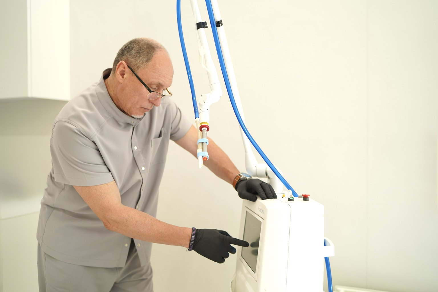
At Sandler Wellness Center (23 Kniaziv Ostrozky St, Kyiv), we conduct a comprehensive dermatoscopic evaluation of nevi using the VIDIX system — this is not just a "visual inspection," but a digital analysis of pigment structures with the ability to track dynamics over time. This approach allows us to distinguish what can be safely removed cosmetically from what requires oncological vigilance.
Next — without embellishments, with specific criteria.
What is a nevus and why not all moles are the same
A nevus is a benign skin formation composed of melanocytes (pigment cells). It is not just a "spot on the skin." Depending on the location of melanocytes in the layers of the dermis, nevi are classified as intradermal, junctional, and compound — and each type has a different risk of transformation.
Most people have between 10 and 40 nevi. This is normal. But there are nuances. Having more than 50 nevi is itself a risk factor for developing melanoma (British Journal of Dermatology study, 2022, n=11,000+). Atypical nevi (dysplastic) — those with irregular borders, heterogeneous coloring, a diameter greater than 6 mm — require mandatory dermatoscopy, not cosmetic removal "just in case."
There are formations that are not nevi at all, although they look similar. Seborrheic keratosis, dermatofibroma, angiofibroma — their removal is technically simple and safe, but only after differential diagnosis. Removing a melanoma, mistaking it for a regular mole — is a classic tragic mistake.
What is important to understand practically: a nevus itself does not hurt, does not itch, does not bleed under normal conditions. If any of these symptoms are present — it is no longer "which mole to remove," it is "when to see an oncologist." The answer: now.
ABCDE Rule — an international standard for the primary evaluation of nevi:
- A (Asymmetry) — asymmetry: the halves do not match
- B (Border) — irregular, blurred, serrated edges
- C (Color) — uneven color, multiple shades within one formation
- D (Diameter) — diameter greater than 6 mm (larger than a pencil eraser)
- E (Evolution) — any change over weeks or months
One positive criterion is already a reason for dermatoscopy. Two or more — an urgent examination by a dermatologist-oncologist.
Which nevi can be safely removed and which cannot: specific criteria
This is the section through which people search for the article. I will give a direct answer.
It is safe to remove (after mandatory dermatoscopy):
- Intradermal nevi — dome-shaped, soft, uniform in color, without signs of atypia. They are removed with laser or radiofrequency with a good cosmetic result.
- Nevi in areas of constant trauma (belt, strap, collar) — but only after confirming the absence of atypia. Chronic trauma theoretically increases the risk of transformation, and this is a real medical indication for removal.
- Seborrheic keratoses that resemble moles — after differential diagnosis.
- Nevi that are cosmetically bothersome, provided their benign nature is confirmed.
Do not remove without prior oncological consultation:
- Formation with any ABCDE sign
- New formation that appeared after age 40
- Any formation that has changed in the last 3 months
- Formation that bleeds or has crusted over
- Mole after sunburn in this area
- Formation on palms, soles, nail bed (acral localization — increased risk)
- Formation with confirmed dermatoscopically benign structure — removal is safe
I see a recurring situation in practice: a patient comes to remove "this little mole," and under the dermatoscope, we find an atypical nevus with irregular streaks and regression areas. We proceed with removal — but already with a different protocol: surgical excision with margins, mandatory histology. No laser, no radio knife. Because the diagnosis is more important than cosmetics.
Results and scientific evidence: what early dermatoscopy provides
The numbers here are telling. A Cochrane meta-analysis (2018, 23 studies, n=9,000+) showed that the implementation of systematic dermatoscopy reduces the number of unnecessary removals of benign formations by 42% while simultaneously increasing the detection of melanoma at early stages by 34%.
The 5-year survival rate for melanoma in situ (stage 0) — 99%. At stage I — 92-98%. Stage IV — 15-20%. This is not abstract statistics. This is the difference between 20 minutes of outpatient removal and years of chemoimmunotherapy.
In our practice at Sandler Wellness Center, based on the results of dermatoscopic screening, we redirect approximately 8-12% of formations from all visits for surgical excision — meaning the vast majority are truly benign and removed without oncological risk.
After confirmed removal of a benign formation with laser or radiofrequency, healing takes 7-14 days. Generally, there is no scar left. After surgical excision with margins (in cases of atypia) — a scar is present, but its size depends on the location and technique of suturing. The histological result is usually ready within 7-10 days.
Contraindications and safety of nevus removal
Any removal of a nevus is a medical procedure. Even if it seems like a cosmetic triviality.
Absolute contraindications for laser/radiofrequency removal:
- Suspected malignancy based on dermatoscopy (the method of choice — only surgery with histology)
- Blood coagulation disorders or taking anticoagulants without dose adjustment
- Active herpes infection in the intervention area
- Pregnancy (first trimester — a relative contraindication for most physiotherapeutic effects)
What is important to know before the procedure:
For 2-4 weeks before removal, avoid intense sun exposure in the area. Tanned skin increases the risk of post-inflammatory hyperpigmentation after laser treatment.
After removal: do not wet the wound for the first 24 hours, apply the recommended antiseptic, avoid the sun for at least 4-6 weeks, and be sure to use SPF 50+ on the healing area.
The side effects of removing benign nevi are minimal when following the protocol: slight hyperemia for 2-3 days, crusting for up to 10-14 days. Hypertrophic scars after laser occur in less than 1% of cases with properly selected power and type of laser.
Price and appointment at Sandler Wellness Center, Kyiv
At Sandler Wellness Center at 23 Kniaziv Ostrozky St, Kyiv, a consultation with a dermatologist-oncologist includes a complete dermatoscopic evaluation using the VIDIX system.
Service costs:
| Service | Price |
|---|---|
| Consultation with an oncologist, PhD + VIDIX dermatoscopy | 3,500 UAH |
| Follow-up consultation with an oncologist, PhD + VIDIX dermatoscopy | 1,600 UAH |
| Laser-radiofrequency removal of skin formations | from 1,000 to 30,400 UAH |
| Histological examination | 1,200 UAH |
The cost of removal depends on the number, size, and type of formations — the exact amount is determined after examination. We recommend histological examination for all removed formations without exception: this is the only way to obtain a morphological diagnosis.
You can schedule a consultation through the website sandler.com.ua or by calling the administrator. Appointments are held at Sandler Wellness Center in the center of Kyiv, with convenient transport accessibility.
Prices may change — please confirm the current cost with the administrator or on sandler.com.ua.
Frequently Asked Questions (FAQ)
Q: Is it dangerous if a mole was nicked by a razor or nail?
A: A one-time superficial injury to a benign nevus does not itself cause transformation into melanoma — scientific data does not support this. However, if the damage occurs regularly or the wound does not heal for more than 2-3 weeks, or if there are secretions or color changes — dermatoscopy is necessary. Systematic trauma is a clinical indication for nevus removal, but only after confirming its benignity.
Q: Can a mole be removed using a cream or pencil from the pharmacy?
A: No. No home "removal" product for moles has undergone clinical trials for effectiveness and safety. Acids and coagulating agents destroy tissue without controlled margins and without histological material. If the formation turns out to be atypical, such "treatment" makes proper diagnosis impossible and may accelerate the spread of cells. The only safe option is removal in a medical facility after dermatoscopy.
Q: How much does mole removal cost in Kyiv and what does it include?
A: At Sandler Wellness Center, the cost of a dermatoscopic consultation with an oncologist, PhD, is 3,500 UAH, and a follow-up is 1,600 UAH. Laser-radiofrequency removal of skin formations is calculated from 1,000 to 30,400 UAH depending on the number and characteristics of the formations. Histology is 1,200 UAH separately. The exact amount is determined by the doctor after examination. You can schedule an appointment at sandler.com.ua.
Q: What does melanoma look like at the beginning and how does it differ from a regular mole?
A: Early melanoma often looks like a regular spot that has "slightly changed." Key signs according to the ABCDE system: asymmetry of shape, irregular or blurred edges, multiple shades of color within one formation (brown, black, gray, pink), diameter greater than 6 mm, and most importantly — any dynamics of change. It is impossible to distinguish an atypical nevus from melanoma in situ even for an experienced doctor with the naked eye — this is precisely why dermatoscopy exists.
Q: Is it necessary to perform histology after the removal of each mole?
A: Yes, this is the standard of evidence-based medicine. Histological examination of the removed material is the only way to obtain a morphological diagnosis. Even if dermatoscopy shows a benign structure, histology confirms or clarifies this conclusion. Refusing histology when removing pigmented formations is equivalent to refusing a diagnosis. At Sandler Wellness Center, histology is included in the removal protocol as a recommended but not mandatory service.
Q: How long after removal can I sunbathe or visit the beach?
A: At least 4-6 weeks after laser or radiofrequency removal of any skin formation. Young skin at the healing site is extremely sensitive to UV radiation and reacts with hyperpigmentation — a dark spot that can last for months. Be sure to use SPF 50+ on the area daily, even on cloudy days, throughout the entire season after the procedure.
Conclusion
The question "which moles can be removed" does not have a correct answer without dermatoscopy. This is where safe medicine in this topic begins and ends. What looks like a regular mole may have structures under magnification that are incompatible with the concept of "cosmetic procedure." Conversely, a formation that scares the patient often turns out to be completely benign and can be removed in 10 minutes.
Sandler Wellness Center in Kyiv provides a full cycle: consultation with a dermatologist-oncologist with VIDIX dermatoscopy, clinical decision-making, removal of confirmed benign formations, and histological control of the material. All in one place, according to clear medical protocols.
Schedule an examination through sandler.com.ua or contact the administrator of Sandler Wellness Center at 23 Kniaziv Ostrozky St, Kyiv. A preventive examination once a year is the cheapest and most effective oncological insurance that exists.
 of the brain and neuroprotection at Sandler Wellness Center, [Kyiv](https://blog.sandler.com.ua/персональний-тренер-Kyiv-фітнес-sandler-індивідуальний-підхід-до-вашого-тіла)](https://yplfdyggcmsagjsuajof.supabase.co/storage/v1/object/public/sandler-blog/photobank/breath-003.jpg)