Every year, over 7,000 new cases of melanoma are registered in Ukraine — and about 40% of them are detected at stage III–IV, when the five-year survival rate drops below 20%. Meanwhile, melanoma detected at the in situ stage or T1a is cured in 98–99% of cases (data from the National Cancer Registry of Ukraine, 2023). The difference between these two scenarios often comes down to a simple 15-minute consultation with dermatoscopy.
This is where VIDIX becomes a pivotal tool. It is not just a magnifying glass: a digital dermatoscope with polarized lighting allows us to see structures at a depth of 0.2 mm, unreachable by the eye of even an experienced doctor. In our practice at Sandler Wellness Center — where I have conducted over 3,000 such examinations — every third patient comes with a "safe mole," which appears entirely different on the VIDIX screen.
This article is not meant to scare you. It is about what true melanoma screening looks like from the inside — what exactly the doctor sees, why some signs are alarming while others are not, and when 15 minutes can change the prognosis forever. If you are in Kyiv and have never had your moles checked by a dermatologist-oncologist, this article is for you.
What is VIDIX Dermatoscopy and Why is it More Accurate than Simple Examination
Dermatoscopy is a non-contact microscopic visualization method of the skin with 10–70 times magnification, eliminating surface reflection and allowing for the assessment of subepidermal structures. VIDIX is a specific digital dermatoscopy platform with cloud image archiving, enabling not just a one-time examination of a mole today, but also the ability to compare its dynamics over 6–12 months.
Why is this crucial? The human eye without a dermatoscope distinguishes melanoma from a benign nevus in 60–70% of cases. With a dermatoscope in the hands of an experienced specialist, the accuracy increases to 89–95% (study from the Journal of the American Academy of Dermatology, 2022, n=2,844). VIDIX adds digital archiving to this: even if a formation currently looks neutral, a year later, comparative photos will immediately reveal regressive structures or changes in the pigment pattern.
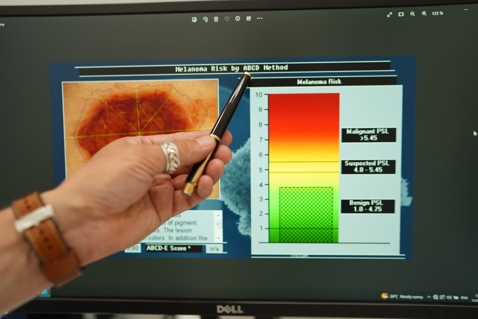
What exactly does the doctor assess under the dermatoscope? There are several analytical systems, but the most commonly used in European practice is the ABCD Stolz algorithm and the 7-point checklist. I use both in parallel because they capture different signs.
Key structures visible in VIDIX:
Pigment network (reticular pattern) — uniform in benign nevi, atypical (thickened, disrupted) in melanoma. Pseudopodia — stalk-like projections at the edge — almost always indicate oncological caution. Regressive zones (whitish scarring) and blue-gray veils indicate tumor invasion of the dermis. Atypical vessels — punctate, irregular, needle-like — appear when melanoma thickness exceeds 0.75 mm.
In our practice at Sandler Wellness Center (23 Kniaziv Ostrozky St, Kyiv), each VIDIX examination includes parallel photo documentation, a protocol, and, if necessary, dynamic monitoring.
Who Needs Dermatoscopy and How the Protocol Works at Sandler Wellness Center
Patients often ask: "I just have moles, why do I need this?" I typically respond: there are more risk factors than most people think.
Checklist for indications for VIDIX dermatoscopy:
- More than 50 melanocytic nevi on the body
- Atypical (dysplastic) nevi in history — personal or family
- Melanoma in first-degree relatives
- Prolonged or intense sun exposure, sunburns before the age of 18
- No examination by a dermatologist-oncologist in the last 12 months
- A mole that has changed color, shape, or size in the last 6 months
- Formation with bleeding, itching, or crusting
- I–II skin phototype (red or light hair, blue eyes)
If you marked at least 2 items — screening is necessary.
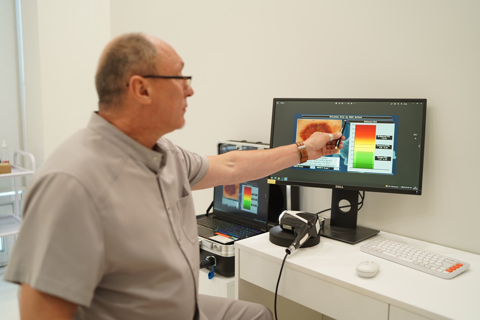
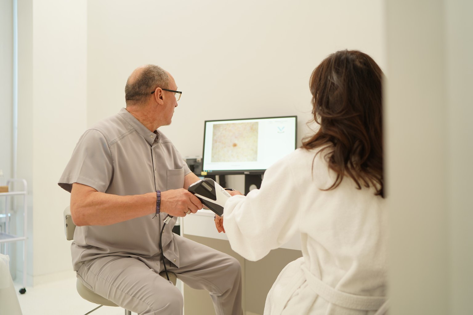
How the appointment works at Sandler Wellness Center. The patient arrives without preparation. The first 5 minutes involve collecting medical history: risk factors, dynamics of changes, complaints. Then I perform body mapping: a systematic examination of all visible nevi, including the scalp and soles. Each suspicious formation is photographed with VIDIX in both polarized and non-polarized modes. On the monitor screen, I immediately explain to the patient what I see.
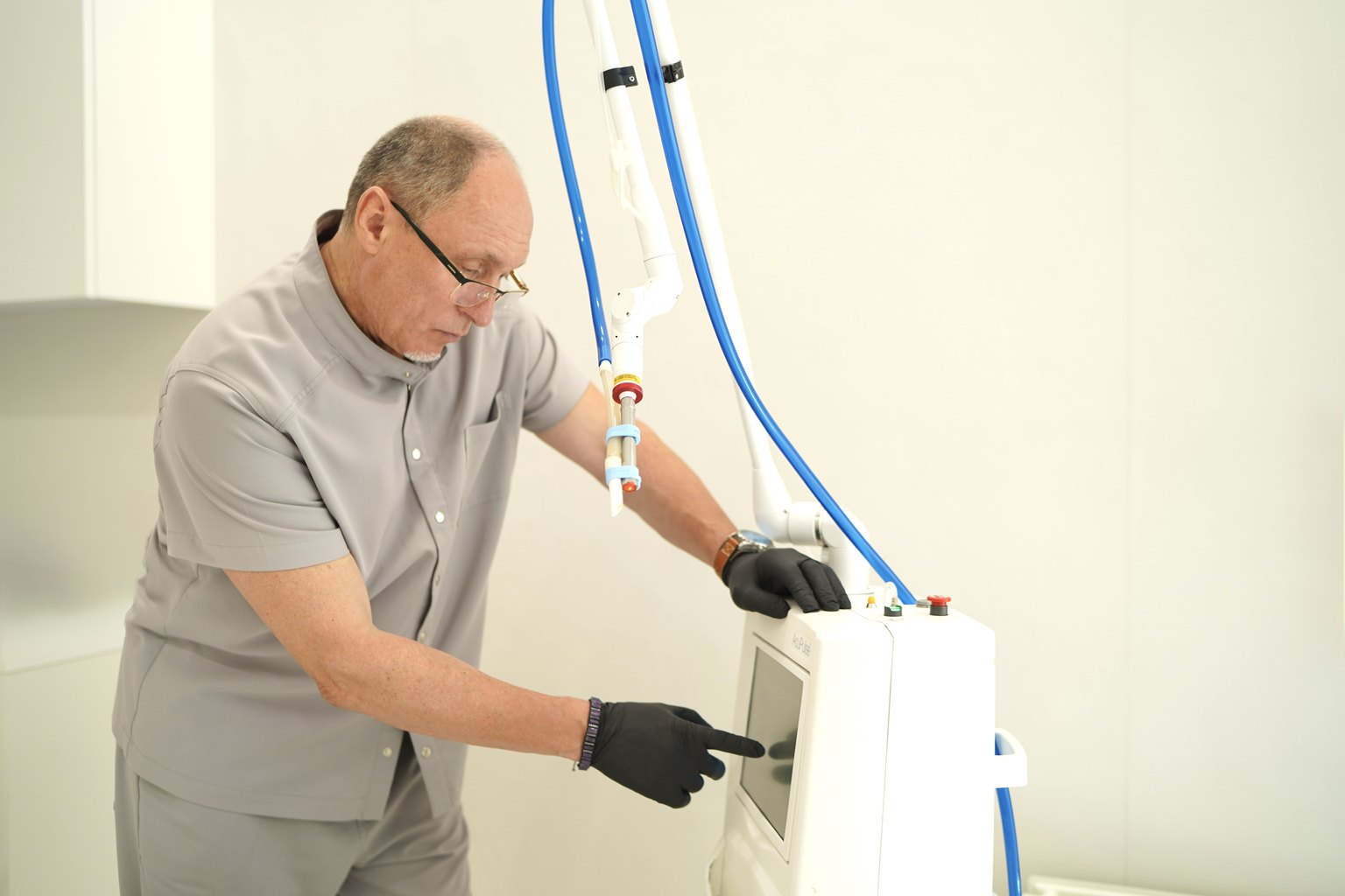
If there are alarming signs — I discuss three scenarios: dynamic monitoring (repeat dermatoscopy in 3 months), excision with histological examination, or laser-radio wave removal. The choice depends on the specific VIDIX picture, localization, and the patient's preferences.
An important detail: VIDIX maintains an image database. During a repeat visit, the program automatically compares the current state with the previous one. A change in area of even 15% or the appearance of new vascular structures is immediately visible in the overlay comparison.
→ See also: Early Stage Melanoma: 5 Signs Only a Dermatoscope Can See
Scientific Evidence and Real Results: What Practice Shows
Dermatoscopy with digital archiving has reduced melanoma mortality in Scandinavian countries by 26% over 10 years of widespread implementation (Acta Dermato-Venereologica, Stockholm, 2021, n=12,400). This is not a theoretical figure — it is the result of a change in screening protocol.
In our practice: from 2023 to 2024 at Sandler Wellness Center during VIDIX screening, we detected 14 melanomas at early stages (T1a–T1b) in patients who came to "just check their moles." The average tumor thickness was 0.4 mm. All 14 cases were radically operated on, none required chemotherapy. Another 23 patients were recommended dynamic monitoring upon detection of moderately atypical nevi — annual repeat examination.
What can you realistically expect after the first visit? If the formation is benign — a conclusion with VIDIX photo and a recommendation for the next examination. If there is atypia of grade I–II — a monitoring protocol. If the picture is alarming — a referral for excision the same day or scheduling surgery within a week. No "come back in 3 months to check" when there are real oncological signs.
Another important nuance: many patients fear that they will "cut out" everything indiscriminately. On the contrary, digital dermatoscopy reduces the number of excisions. I see this every week: a person comes with 5 formations that a surgeon elsewhere wanted to remove "just in case," and 4 of them turn out to be classic Clark nevi without any alarming signs after VIDIX.
Contraindications and Limitations of the Method: What is Important to Know Before the Consultation
VIDIX dermatoscopy is a non-invasive, non-contact method. There are no absolute contraindications.
Several practical limitations that I always discuss with patients.
Formations under the nail plate (subungual) or on mucous membranes are more challenging to assess — VIDIX provides a picture, but it requires more careful interpretation. Formations with ulceration or bleeding also complicate the dermatoscopic picture. Very dark skin (phototype V–VI) has specific features of the pigment pattern, and here an experienced specialist is needed who understands ethnic variations of the norm.
If a mole has already been removed and there are scar changes around — this is not a contraindication, but I ask the patient to bring the previous protocol or histological conclusion if available.
Also: VIDIX is not suitable for diagnosing non-melanocytic skin tumors (basal cell carcinoma, squamous cell carcinoma) in the classic melanoma algorithm — there are separate dermatoscopic criteria for them. But the good news is: I evaluate all these formations within one appointment.
Price and Appointment for VIDIX Dermatoscopy at Sandler Wellness Center, Kyiv
Sandler Wellness Center is located at 23 Kniaziv Ostrozky St, Kyiv. Appointments with a dermatologist-oncologist are by prior arrangement, without queues.
Cost of dermatological-oncological services:
| Service | Price |
|---|---|
| Consultation with an oncologist, PhD + VIDIX dermatoscopy | 3,500 UAH |
| Follow-up consultation with an oncologist, PhD + VIDIX dermatoscopy | 1,600 UAH |
| Histological examination (after excision) | 1,200 UAH |
| Laser-radio wave removal of skin formations | from 1,000 to 30,400 UAH |
A full consultation includes: medical history collection, body mapping, digital VIDIX dermatoscopy of all suspicious formations, photo archiving, written examination protocol, and recommendations for further tactics. A follow-up consultation is a comparative analysis of VIDIX photos from the previous visit: this is where changes that a one-time examination might miss are revealed.
If removal is needed based on the examination results, the cost of the laser-radio wave procedure is specified individually — it depends on the size, number, and localization of the formations.
To make an appointment, call sandler.com.ua or contact the center administrator directly.
Prices may change — please check the current cost with the administrator or on sandler.com.ua.
→ See also: ABCDE Rule: How to Check a Suspicious Mole in 2 Minutes
Frequently Asked Questions about VIDIX Dermatoscopy
Q: How often should I undergo dermatoscopy if my moles do not change externally?
A: If you have no risk factors (fewer than 50 nevi, no atypical formations, no family history of melanoma), once a year is sufficient. Patients with dysplastic nevus syndrome or a history of melanoma are recommended to have an examination every 6 months at Sandler Wellness Center. Frequency is determined individually based on the results of the first VIDIX screening — and this is documented in the protocol.
Q: Is VIDIX dermatoscopy painful? Is preparation needed?
A: The procedure is completely painless and non-contact — the device does not touch the skin. No preparation is needed: just come without makeup on your face if there are formations there. I recommend not painting your nails (if there are formations in the subungual area) and wearing comfortable clothing that is easy to remove for body examination. The entire procedure takes 15–30 minutes depending on the number of formations.
Q: Can VIDIX replace a biopsy and provide a definitive diagnosis of skin cancer?
A: No, and this is fundamental. VIDIX is a tool for selection and risk stratification, not a substitute for histopathology. If the dermatoscopic picture raises suspicion of melanoma, the only reliable diagnosis is histological examination of the tissue after excision. VIDIX determines which formation to remove first and with what margins — this is clinically very important information.
Q: I am 25 years old and have no health issues. Why should I check my moles now?
A: This is precisely the point of screening. The median age at the time of melanoma diagnosis in Ukraine is 47 years, but 15–20% of cases are detected in people under 40 (data from the National Cancer Registry, 2022). Melanoma develops over the years. If you come in at 25 and establish a base of VIDIX photos, then a year later, comparative analysis will immediately show any changes — even before they become visible to the eye.
Q: Does VIDIX only detect melanoma, or does it also detect basal cell carcinoma and squamous cell carcinoma?
A: VIDIX as a platform — yes, as a tool it allows for the assessment of any skin formation. Basal cell carcinoma, squamous cell carcinoma, dermatofibroma, angiomas — all have characteristic dermatoscopic signs that an experienced specialist recognizes. Therefore, it is more accurate to say "screen for skin formations" rather than "check moles" — this is a broader and more precise approach to oncological prevention.
Q: Can dermatoscopy be performed on a child?
A: Yes. In children, dermatoscopy is performed according to the same principles. Congenital nevi (congenital moles over 1.5 cm) require mandatory VIDIX monitoring from birth — the risk of malignancy in large congenital nevi reaches 4–6% over a lifetime (Pediatric Dermatology, 2021). At Sandler Wellness Center, I accept patients from the age of 5.
Conclusion: 15 Minutes Worth Finding
VIDIX dermatoscopy is not excessive medical caution. It is a tool that separates real risk from imagined, without causing any discomfort to the patient. I see the result every week: a person leaves the office either with peace of mind ("your moles are benign, the next examination is in a year") or with a specific action plan while the situation is still fully controllable.
Melanoma detected in time is not a death sentence. Missed melanoma due to delayed examination is an entirely different statistic.
You can schedule a consultation with an oncologist, PhD, with VIDIX dermatoscopy at Sandler Wellness Center by visiting sandler.com.ua or by contacting the administrator at 23 Kniaziv Ostrozky St, Kyiv. Appointments are made by schedule, without queues.
Do not postpone what can be resolved in 15 minutes.